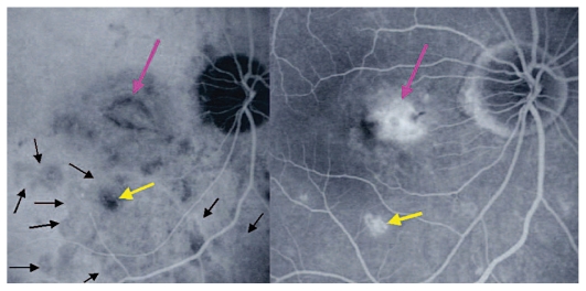
Figure 2D.
Choroidal neovascularization complicating MFC shown on ICGA (left) and FA (right). As shown in figure 2B, choriocapillaris ischemia is usually extensive in MFC and can only be detected by ICGA. This figure shows the development of CNV in the right eye (crimson arrow) concomitant with an acute recurrence of MFC causing extensive choriocapillaris ischemia (black arrows) not visible on FA. The darker areas are old cicatricial foci that can also be seen on FA (window defects, yellow arrows). (Courtesy of Alessandro Mantovani, Como, Italy)

