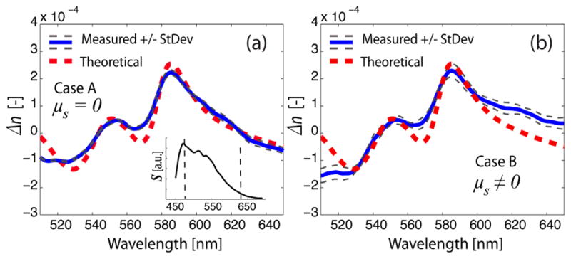
Fig. 2.

Measured change in the real part of the refractive index of the Hb phantoms without scattering (case A) (a), and with scattering (case B) (b). The theoretical change in the real part of the refractive index of Hb, with a concentration of 40 g/L, is also plotted. The inset illustrates the source’s spectrum, S, with the dotted lines denoting the bandwidth used for estimating concentration using the real part of the RI.
