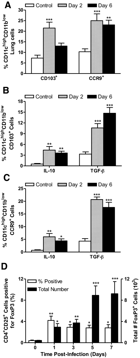
Figure 7. Ft induces the development of pulmonary tDCs and Tregs.
Two groups of C57BL/6 mice were infected with 1 × 10³ CFU of BHI-grown Ft LVS and killed at Days 2 and 6 PI. One group of sham-inoculated mice served as a control. (A) CD11chigh lung cells were evaluated by flow cytometry for surface expression of the tDC markers, CD103 and CCR9. CD11chighCD103+ cells (B) and CD11chighCCR9+ cells (C) were analyzed for IL-10 and TGF-β cytokine production by flow cytometry. (D) Tregs were identified on the basis of FoxP3 expression. Gating on CD4+ cells, the percentages, and total numbers of CD25+FoxP3+ cells were calculated. The values are expressed as mean ± sem from four independent experiments (n=14 total mice/group). *P < 0.05; **P < 0.01; and ***P < 0.001.

