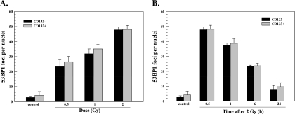
Figure 3.
53BP1 foci after irradiation of NSC11 cells in vitro. Monolayer cultures of CD133- cells (black bars) or CD133+ cells (gray bars) were irradiated and analyzed 0.5 hours later (A) or irradiated with 2 Gy and analyzed at the specified times (B). Values shown represent the mean ± SD of three independent experiments in which 25 cells were scored. The 2-Gy/0.5-h value is repeated in A and B.

