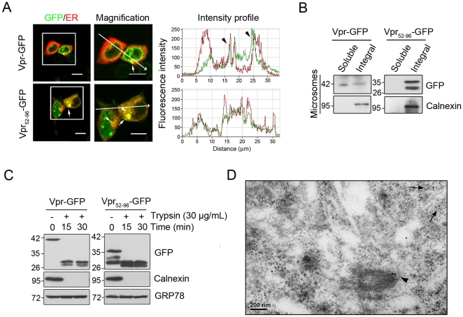
Figure 2. Transport of Vpr to the ER.
A, Confocal immunofluorescent microscopy localized Vpr at the ER. ER was labeled using plasmid encoding ER-targeted Discosoma red fluorescent protein (DsRed-ER). Yellow fluorescence indicates the overlapped regions between Vpr and ER (white arrow). Bar: 10 µm. B, Treatment with 100 mM sodium carbonate confirmed that Vpr and the ER integral protein Calnexin were present in the alkaline-resistant fraction. C, Isolated microsomal fractions treated with trypsin indicate that Vpr inserted into the ER by the C-terminal TMD. D, Using immunogold electron microscopy, Vpr particles (15 nm) were shown to be located on the membrane of mitochondria (arrowhead) and ER (arrow). These results are representative of three independent experiments.

