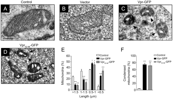
Figure 5. Vpr protein influences the morphology of mitochondria and integrity of MOM.
A, Electron microscopic analysis of HEK293 cells showed that mitochondria were intact with a double-layer membrane and regular arrangement of cristae. Bar: 200 nm. B, Similar to wild-type cells, expression of GFP did not influence the morphology of the mitochondria. Bar: 200 nm. C, Following Vpr-GFP expression, marked changes in the architecture of the cristae was observed. Vpr led to swollen cristae, condensed matrix (arrowhead) and gradually disappearance of outer membrane (arrow). Bar: 200 nm. D, The same phenomenon was observed with Vpr52–96-GFP expressing cells. Mitochondria were condensed with swollen cristae. Bar: 200 nm. E, Based on TEM images, Vpr induced mitochondrial fragmentation and the length of mitochondria was evidently shorter. Twenty five cells were scored in three independent experiments with means ± S.D. F, Vpr also resulted in the condensation of mitochondria. For panels E and F, 25 cells were scored in three independent experiments with means ± S.D. *** indicates statistically significant difference (p<0.001) between the control and Vpr-expressing cells.

