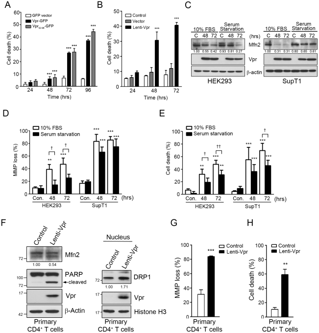
Figure 8. Vpr-mediated mitochondrial damage causes cell death in HEK293 and CD4+ T cells under normal growth condition or serum starvation.
A, HEK293 cells were transfected with GFP vector the plasmid encoding Vpr-GFP or Vpr52–96-GFP, and harvested at different time (hours) post-transfection for PI staining. The percentage of dead cells among GFP-expressing cells was determined by flow cytometry. *** (p<0.001) indicates significantly different from the GFP vector control. B, HEK293 cells were infected with lenti-vector (control) or Lenti-Vpr and harvested at different time (hours) post-infection for PI staining. The percentage of dead cells was determined by flow cytometry. *** (p<0.001) indicates significantly different from the control. C, HEK293 and SupT1 cells were grown in 10% FBS or starved for 24 hours, and infected with Vpr-expressing lentivirus for 48 and 72 hours. The expression of Mfn2 was decreased in serum-starved HEK293 or SupT1 cells. The quantitative expression of Mfn2 was measured by Image J and normalized with the expression of β-actin. C indicates Vpr negative lentiviral control. D, MMP loss was determined after Vpr-expressing lentivirus infection. Vpr significantly impaired MMP in serum-starved HEK293 and SupT1 cells. E, Vpr expression led to cell death in serum-starved HEK293 and SupT1 cells. For panels D and E, results are the means ± S.D. of three independent experiments. * (p<0.05), ** (p<0.01) and ** (p<0.001) indicate significantly higher than the Vpr negative lentiviral control (Con.). † (p<0.05) and †† (p<0.01) indicate significantly different between 10% FBS and serum starvation. F, Human primary CD4+ T cells were isolated from peripheral blood mononuclear cell (PBMC) and infected with Vpr-expressing lentivirus for 72 hours. The expression of Mfn2 was decreased and the expression of nuclear DRP1 was increased in human primary CD4+ cells. The relative expression levels of Mfn2 and DRP1 were measured by Image J and normalized with the expression of β-actin. G, MMP loss was determined after Vpr-expressing lentivirus infection. Vpr led to a significant MMP loss in human primary CD4+ T cells. H, Vpr expression led to cell death in human primary CD4+ T cells. For panels G and H, results are the means ± S.D. of three independent experiments. ** (p<0.01) and *** (p<0.001) indicate significantly higher than control human primary CD4+ T cells. Band intensities were calculated using Image J. Relative intensities are shown at the bottom of each panel.

