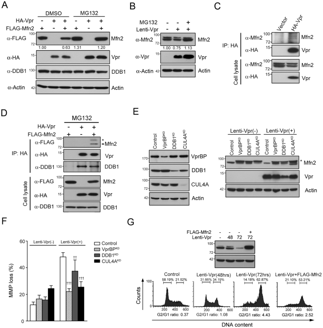
Figure 10. Vpr downregulated Mfn2 expression via VprBP-DDB1-CUL4A ubiquitin ligase complex.
A, HEK293 cells were transfected respectively with plasmids encoding HA-Vpr and Flag-Mfn2 for 32 hours and treated with proteasome inhibitor MG132 (5 µM) for 16 hours. The expression of Mfn2 was recovered after MG132 treatment. B, HEK293 cells were infected with Lenti-Vpr for 56 hours and treated with proteasome inhibitor MG132 (5 µM) for 16 hours. The expression of Mfn2 was not decreased after MG132 treatment. C, HEK293 cells were transfected with the plasmid encoding HA-Vpr and harvested after 48 hrs. Cell lysates were immunoprecipitated with anti-HA antibody, and detected by Western blotting. D, HEK293 cells were transfected respectively with plasmids encoding HA-Vpr and Flag-Mfn2 for 32 hours and treated with MG132 (5 µM) for 16 hours. Cell lysates were immunoprecipitated with anti-HA antibodies, and the coimmunoprecipitated proteins were detected by Western blotting. * indicates the ubiquitinated Mfn2. E, HEK293 cells were silenced by shRNA against DDB1, VprBP or CUL4A, and infected with lentivirus carrying Vpr for 72 hours. The ubiquitinated Mfn2 was detected in VprBPKD, DDB1KD, CUL4AKD cells. Moreover, Vpr infection did not decrease Mfn2 expression in VprBPKD, DDB1KD, CUL4AKD cells. * indicates the ubiquitinated Mfn2. F, The MMP loss was determined by flow cytometry after Lenti-Vpr infection. Knockdown of DDB1, VprBP and CUL4A reduced the percentage of MMP loss after Lenti-Vpr infection. G, HEK293 cells were infected with Lenti-Vpr for 24 hours and transfected with the plasmid encoding FLAG-Mfn2 for 48 hours. Cells were harvested and analyzed by Western blotting and flow cytometry. Band intensities were calculated using Image J. Relative intensities are shown at the bottom of each panel.

