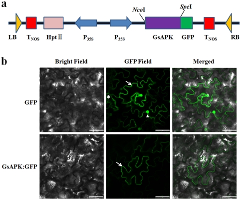
Figure 4. GsAPK protein targets to the plasma membrane of N.benthamiana plant cells.
a. Schematic representation of construct used for subcellular localization of the GsAPK protein. b. Subcellular localization assay of the GsAPK:GFP fusion protein in N. benthamiana epidermal cells. Micrographs showing cells expressing GFP (control, upper lane) or GsAPK:GFP (bottom lane) fusion protein, which were examined under bright-field illumination (left), and under fluorescent-field illumination (middle) to examine GFP fluorescence, and by confocal microscopy (right) for an overlay of bright and fluorescent illumination. Scale bar, 50 µm. Arrow, plasma membrane; asterisk, cytosol; triangle, nucleus.

