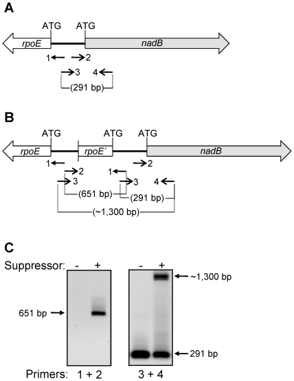
Figure 4. Identification of the genetic rearrangement in the suppressor strain.
PCR amplifications were carried out to narrow down the site of possible genetic rearrangement in the rpoE-nadB region of the chromosome. (A, B) Approximate drawings showing the rpoE and nadB genes from the wild-type (A) and suppressor (B) strains, as well as the positions of primers (numbered 1 to 4), their orientations, and size of the amplified DNA fragments. (C) Agarose gels showing the results of PCR amplifications from the wild-type and suppressor strains. Numbers 1 to 4 refer to the primers shown in (A) and (B).

