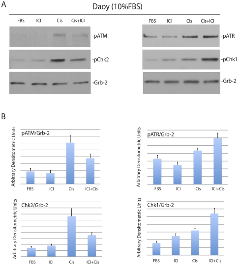
Figure 4. Inhibition of ERβ modulates cisplatin-induced phosphorylation of cell cycle checkpoint proteins.
Panel A: Western blot analyses showing levels of the phosphorylated ATM, ATR, Chk1 and Chk2 in constitutively growing Daoy cells (10%FBS) treated with cisplatin (1 µg/ml) in the presence (Cis+ICI) or absence (Cis) of ICI182,780 (10 µM). The cells without treatment (FBS), or cells treated with ICI182,780 only (ICI) were used as controls. Panel B: Densitometry of Western blots depicted in Panel A evaluated by EZQuant-Gel 2.17 software. Levels of pATM, pATR, pChk1 and pChk2, were normalized with the corresponding levels of Grb-2. Data represent averages obtained from densitometric measurements of 3 blots with standard deviation and each band was normalized with corresponding loading control, Grb-2.

