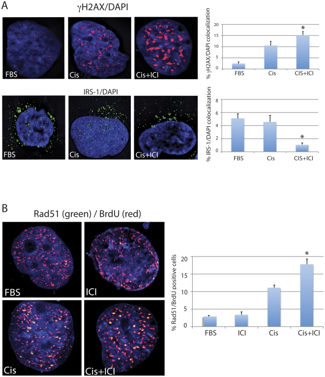
Figure 6. Inhibition of ERβ activates recruitment of Rad51 during S phase DNA repair.
Panel A: Fluorescent images of Daoy cells immunolabeled with anti-histone γH2AX (upper panel) and anti-IRS-1 (lower panel) antibodies. The nuclei are visualized by DAPI staining (blue fluorescence). The histograms represent quantification of the co-localization between γH2AX and DAPI; IRS-1 and DAPI. The data represent average percentage of nuclear voxels (3-D pixels) of γH2AX (red fluorescence) and IRS-1 (green fluorescence) calculated from three independent experiments (n = 3) in which ten randomly selected cells have been evaluated by the Mask analysis included in SlideBook 5 deconvolution software. * indicates value statistically different from the sample labeled Cis (paired student t-test; P≤0.05). Panel B: Fluorescent images of the cells labeled with anti-Rad51 (green fluorescence) and with anti-BrdU (red fluorescence) antibodies. Exponentially growing cultures of Daoy cells (10%FBS) were exposed for one hour to bromodeoxyuridine (BrdU) during the 6 hours treatment with cisplatin (1 µg/ml) in the absence (Cis) or in the presence of 10 µM ICI182,780 (Cis+ICI). The histogram represents quantification of Rad51 positive cells in which Rad51 nuclear foci co-localize with BrdU-labeled DNA. Note, almost 40% increase in the number of cells utilizing Rad51 to repair cisplatin-induced DNA damage (during DNA replication) when the cisplatin treatment is accompanied by ICI182,780. * indicates value statistically different from the sample labeled Cis (paired student t-test; P≤0.05).

