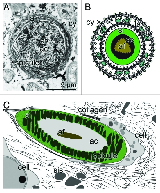
Figure 2.
Demosponge siliceous spicule formation; appositional lamellar growth. (A) During the radial growth of the spicules an organic cylinder (> < cy) is formed into which both silicatein and bio-silica is deposited. In the center an axial filament (af) is located that is harbored by the axial canal (ac). Cross section that had been contrasted with immunogold labeled anti-silicatein antibodies; TEM. (B) Appositional telescopic arrangement of radial, organic cylinders (cy) which become increasingly filled with bio-silica (si), formed form ortho-silicate and the polymerizing enzyme silicatein. In the center is the axial filament (af) located in the axial canal (ac). (C) Schematic section through sponge tissue showing the cut though spicule and the surrounding silica envelope (si). In the center the axial filament (af) and the axial canal (ac) is located. Around the spicule the sclerocytes are arranged, that release the silicasomes (sis) and also collagen.

