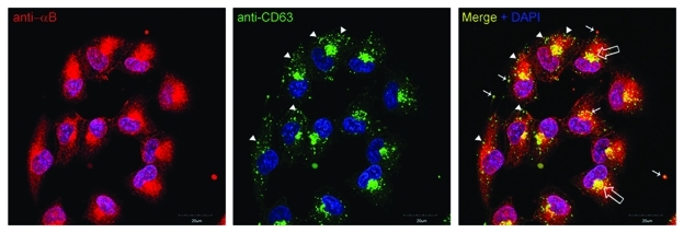
Figure 1.
A confocal z-section (2.5 µm depth) of ARPE19 cells labeled with anti-αB (red) and tetraspanin (CD63) (green). Note co-localization in the perinuclear Golgi (open arrows) and vesicular staining (yellow, thin arrows).Interestingly, the co-localization seems to be more prevalent nearer the perinuclear Golgi (indicated by thin arrows). Note that the green (CD63) and Red (αB) do not go together in the periphery of the cells (arrow heads). Not all vesicles labeled with anti CD63 label with anti αB, indicating that the role of CD63 in αB-containing exosomes may be either transitory or selective. Nuclei are stained with DAPI (blue); scale bar = 20 µm.

