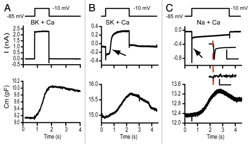
Figure 1.
The dual sine wave technology allows for monitoring capacitance concomitantly with the occurence of voltage or calcium-dependent currents. In each frame the upper panel depicts the stimulus, the middle panel the extracted currents and the lower panels the capacitance measurements. The sine waves are not shown on the stimulus but had frequencies of 6.3 and 9.4kHz. Stimulus artifacts were digitally removed and data filtered with a fourier filter to remove high frequency components. Both (A and B) were recordings from turtle hair cells while (C) was from an immature rat hair cell. (A) illustrates capacitance changes measured with a potassium based intracellular solution where the BK calcium activated potassium current activates rapidly in response to a depolarization to -10 mV. (B) Similar records with a cesium based intracellular solution without the presence of apamin externally. The large calcium current activated the SK calcium activated current, indicated by the arrow. SK activation had no effect on capacitance measurements (depolarization to -10 mV). (C) presents capacitance measurements from a postnatal day 8 rat inner hair cell that transiently expressed Na currents. This rapidly activating, rapidly inactivating current, indicated by arrow, also had little effect on the measured capacitance response. The inset expands in time each response with the scale bars reflecting 10 ms and 100 fF and 0.5 nA. The dashed line indicates onset time so that stimulus artifact can be separated from Na current. A and B were post filtered, while C was not in order to not lose an fast components.

