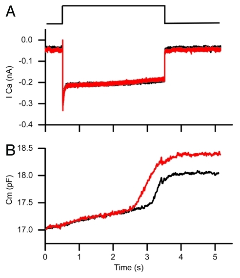
Figure 3.
The dual sine technique demonstrates a major flaw with techniques that require repetitive stimulation in that responses are altered by previous stimulations. The black trace was obtained 10 min after breakthrough and is a response to a depolarization to 75% pf the peak calcium current. The red trace was obtained 5 min later and shows an increased release with insignificant change in the calcium current. This likely reflects a redistribution of vesicles following the first stimulation. The red trace was offset in the y-axis in order to clearly overlay traces.

