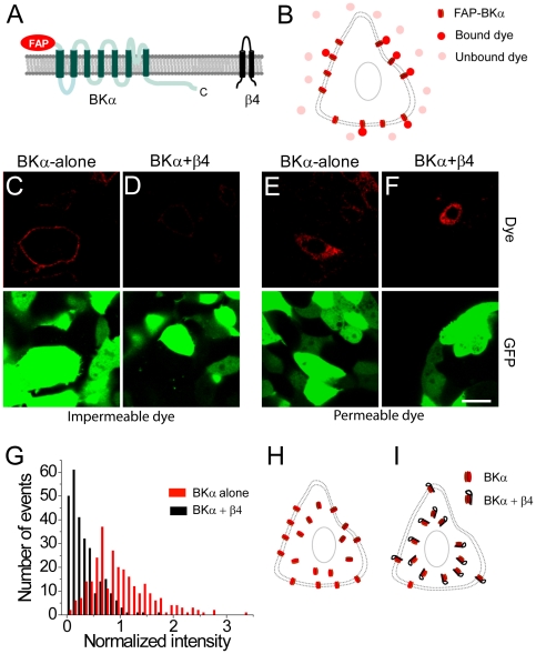
Figure 1. Co-expression of the β4 subunit reduces cell-surface trafficking of BK channels.
(A) Membrane topology of the FAP-BKα and β4 proteins. The FAP tag is at the extracellular, N-terminus. The C-terminus of the BKα subunit is indicated. (B) Schematic of binding of cell-impermeable dye (pink) to the FAP results in significant increase in fluorescence (red). (C) Application of cell-impermeable dye labels only surface BKα channels in live HEK-293 cells co-transfected with FAP-BKα (red) and GFP (green). (D) Same as (C) but in cells co-transfected with β4, FAP-BKα, and GFP showing reduced surface expressed of the FAP-BKα. (E) Application of cell-permeable dye labels intracellular stores of channel in cells transfected with FAP-BKα (red) and GFP (green). (F) Same as (E) but in cells transfected with β4, FAP-BKα, and GFP. Scale bar = 20 µm (C–F). (G) Distribution of surface fluorescence intensity values after application of cell-impermeable dye in transfected cells for FAP-BKα (red bars) or FAP-BKα+β4 (black bars). (H–I) Proposed model for surface distribution of BK channels in the absence (H) or presence (I) of the β4 subunit.

