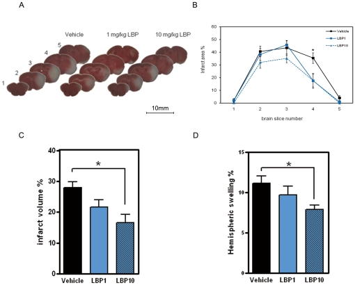
Figure 1. Decreased infarct size and hemispheric swelling in LBP-treated brains after MCAO.
(A) Representative photographs of coronal brain slices stained with 2% TTC. Slice 1, most rostral; slice 5, most caudal. Note the smaller white regions indicating reduced infarct areas in LBP-treated brains. Scale bar = 10 mm. LBP-treated brains showed significantly decreased infarct area % (B), infarct volume % (C) and hemispheric swelling % (D) when compared with vehicle-treated brains. *P<0.05, ANOVA followed by Bonferroni's test, n = 7 to 8 for all groups.

