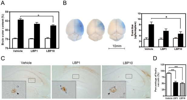
Figure 3. Reduced water content and blood-brain barrier (BBB) disruption in LBP-treated cerebral hemispheres.
(A) Water content in vehicle and LBP-treated cerebral hemispheres 22 hours after reperfusion. White bars, contralateral hemisphere; filled bars, ipsilateral hemisphere. *P<0.05, ANOVA followed by Bonferroni's test, n = 7 each group. (B) Representative photographs of brains after Evans blue (EB) extravasation assay (left). Scale bar = 10 mm. Leakage of EB (blue area) indicated BBB breakdown after MCAO. LBP-treated ipsilateral hemisphere showed decreased EB extravasation (right). White bars, contralateral hemisphere; filled bars, ipsilateral hemisphere. *P<0.05, ANOVA followed by Bonferroni's test, n = 5 each group. (C) Representative IgG IHC showing leaky blood vessels in ipsilateral penumbral areas (interaural 1.98 mm). IgG signal leaked outside the blood vessel lumen (arrow head) in vehicle-treated brain. In LBP-treated brains, the outline of blood vessels was mostly intact and the IgG signal was present inside the vessel lumen. Inserts, higher magnification of typical blood vessels. Scale bar = 200 µm, inserts scale bar = 25 µm. (D) Quantification of blood vessel leakage in ipsilateral penumbral areas. Fewer leaky vessels were observed in LBP-treated brain when compared with the vehicle group. ***P<0.001, ANOVA followed by Bonferroni's test, n = 5 each group.

