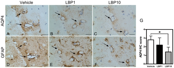
Figure 5. Decreased immunoreactivity of AQP4 in LBP-treated brain.
(A–C) AQP4 IHC signals in swollen end-feet of astrocytes around cerebral vessels in ipsilateral penumbral areas (interaural 1.98 mm). Note the intense AQP4 staining in vehicle-treated vessels after MCAO (arrows, A), which was decreased in both LBP groups (B&C). (D–F) GFAP IHC using adjacent section to AQP4 IHC. Note the GFAP immunoreactivity located around the same cerebral vessels as in the AQP4 immunoreactivity (arrows). Scale bar = 25 µm. (G) Semi-quantification of immunoreactivity of AQP4. *P<0.05, Kruskal-Wallis followed by Dunn's multiple comparison test, n = 5 each group.

