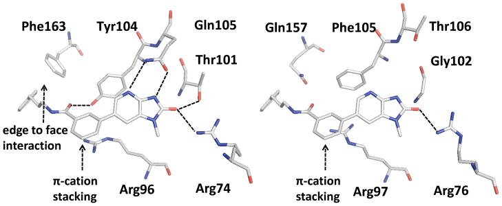
Figure 5.
Comparison of the active sites of PaTMK (left) and hTMK (right). Amino acids associated with the interaction of 46 in PaTMK and their corresponding amino acids in hTMK are represented as sticks with atom type color. The structure of 46 and PaTMK was obtained from the docking study with the X-ray co-crystal structure of PaTMK complexed with 17. The structure of 46 and hTMK was obtained through structure based multiple sequence alignment followed by merging the coordinate of 46 to hTMK structure (PDB ID: 1E2F) from PaTMK structure. H-bond interactions are highlighted with dashed black lines. Edge to face hydrophobic interactions and π-cation stacking are annotated with black arrows.

