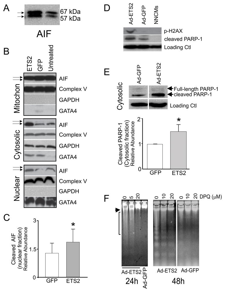
Figure 5. ETS2 over-expression promotes programmed necrosis in cardiomyocytes.
(A) CMs infected with Ad-ETS2 contain two forms of AIF (apoptosis inducible factor): 67kD full-lengh and 57kD cleaved (active). (B) Mitochondrial, cytoplasmic and nuclear enrichment analyses of AIF in untreated CMs or cells infected with Ad-ETS2 or Ad-GFP. Fractionation experiments show that Ad-ETS2 overexpression causes cleaved AIF to translocate from mitochondrial to cytosolic and nuclear compartments. Fractionation enrichment was verified by GATA4 (nuclear), Complex V (mitochondria) and GAPDH (Cytosol). Some mitochondrial associated Complex V was, however, present in each fraction. (C) Relative abundance of AIF in the nuclear fraction of Ad-ETS2 infected NNCMs, after normalization to take into account complex V/mitochondrial contaminants (*t-test, p=0.02, n=4). (D) Ad-ETS2 promotes the release of cleaved PARP-1 and phosphorylation of H2AX in NNCMs. (E) From fractionation studies and after normalization for complex V/mitochondrial contaminants, cleaved PARP-1 abundance significantly increased in the cytosolic fraction after Ad-ETS infection. (F) DPQ treatment completely abolished DNA damage associated with ETS2 overexpression. Overnight pre-incubations prior to Ad-ETS2 infection were performed, and 20 μM DPQ completely abolished all high molecular weight DNA damage 24 and 48 hours post-infection.

