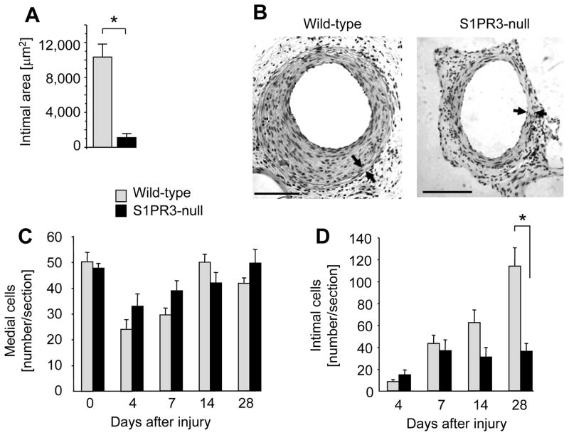
Figure 2. S1PR3 promotes intimal hyperplasia.
Wild-type (wt) and S1PR3-null iliac-femoral arteries were denuded and animals sacrificed at the indicated time points. Arteries were perfusion-fixed and tissue sections stained with H&E. (A) For the 28 day time point, intimal areas were measured using NIH Image software. Data are shown as mean +/− SEM for wt (N=9) and S1PR3-null (N=6). *P<0.001 (B) A typical cross-section of wild-type and S1PR3-null arteries at 28 days after injury is shown. Arrowheads mark the external and internal elastic lamina. The bar indicates 100 μm. The number of cells in media (C) and intima (D) at was quantitated by counting multiple sections that were 250 μm apart. Data (mean +/− SEM, N=7–13 animals per group) are presented as number of cells counted per section. *P<0.05

