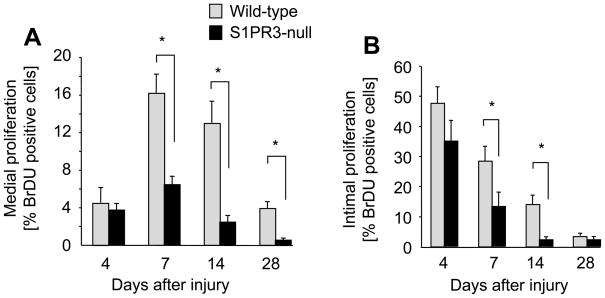
Figure 3. Injury-induced proliferation is increased in wild-type arteries compared to S1PR3-null arteries.
Wild-type (wt) and S1PR3-null iliac-femoral arteries were denuded and injected with BrdU (30 μg/g body weight) at 17, 8 and 1 hour prior to sacrifice at the indicated time points. Arteries were perfusion-fixed and tissue sections stained with anti-BrdU (Roche). The number of positive cells in media (A) and intima (B) was quantitated by counting multiple sections that were 250 μm apart. Data (mean +/− SEM, N=7–13 animals per group) are presented as percent BrdU positive cells. *P<0.05

