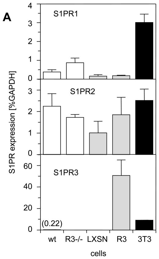
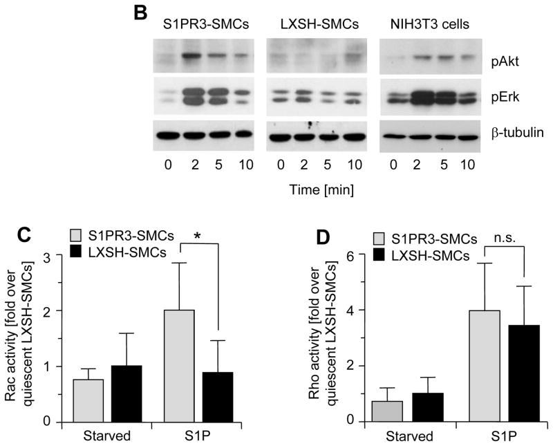
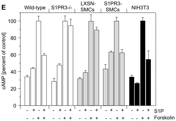
Figure 4. Expression of S1PR3 is linked to S1P-induced activation of Rac1, Akt and Erk, but does not affect S1P-dependent activation of RhoA.
(A) Total RNA was prepared from carotid wild-type SMCs (wt), S1PR3-null SMCs (R−/−), LXSN-SMCs (LXSN, two clones), S1PR3-SMCs (R3, two clones) as well as from NIH3T3 cells (3T3) and analyzed for expression of S1PRs by real time PCR. Data (mean +/− SEM) are presented as percent of GAPDH expression. N=8 for both SMC types, N=4 for LXSN-SMCs, N=6 for S1PR3-SMCs, N=2 for NIH3T3 cells. Following stimulation of LXSH-SMCs (2 clones) and S1PR3-SMCs (2 clones) or NIH3T3 cells with S1P (1 μmol/L) or PDGF-BB (10 ng/mL) as indicated, cells were processed for Western blotting with antibodies against the phospho-forms of Akt and Erk as well as for measuring activities of Rac and Rho. (B) Typical blots for phospho-Akt and phospho-Erk are shown from one out of four experiments that yielded identical results. Blots were reprobed for β-tubulin to demonstrate equal loading. (C) Data for Rac activation were normalized to activity in quiescent LXSH-SMCs and are presented as mean +/− S.D. (N=5) *P<0.05 (D) Data for Rho activation were normalized to activity in quiescent LXSH-SMCs and are presented as mean +/− S.D. (N=4); n.s.=non significant (E) Cells were stimulated for 5 min with S1P (1 μmol/L) and forskolin (3 μmol/L) as indicated. Cyclic AMP was measured and data (mean +/− S.D.) expressed as percent of control (forskolin). The assay was performed in triplicates.


