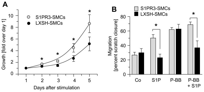
Figure 5. S1PR3 promotes SMC proliferation and migration.
(A). Growth curves of LXSH-SMCs (N=9, 5 different clones) and S1PR3-SMCs (N=11, 2 different clones) were measured using MTT assay. Data (mean +/− SEM, N=11 for S1PR3-SMCs, N=9 for LXSH-SMCs) are expressed as fold absorption at 560 nm over day 1. *P< 0.05 (B) Migration of S1PR3-SMCs (N=6, 2 clones) and LXSH-SMCs (N=6, 2 clones) was measured in the absence or presence of 1 μmol/L S1P, 10 ng/ml PDGF-BB or both using the scratch assay. Data (mean +/− SEM) are presented as percent of “wound” closure. *P<0.05

