
Figure 1.
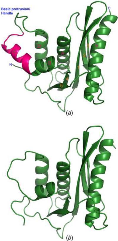
Crystal structures of the full-length XMRV RNase H and its deletion variant ΔC2. (A) Ribbon representation of the overall three-dimensional structure of full-length RNase H. Helix C is highlighted in hot pink, the other parts of the structure in green. Identification of the secondary structure elements (used throughout the manuscript) follows the precedent from related structures. (B) Overall structure of the deletion variant ΔC2 of XMRV RNase H in which helix C was removed. The model is oriented in the same way as in panel A.