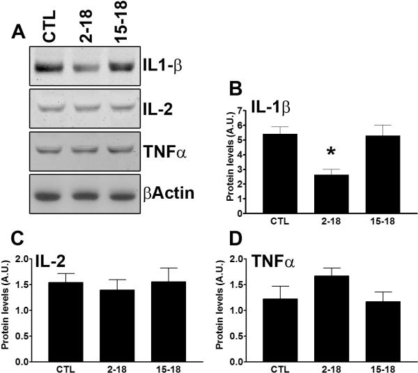
Figure 6. IL-1β levels were reduced in the hippocampus of the Rapa2–18 mice.
Hippocampi from frozen hemi-brains were removed and processed for protein extraction. (A) Representative Western blots from proteins isolated from the hippocampi of CTL, Rapa2–18 and Rapa15–18 mice and probed with the indicated antibodies. (B–D) Quantitative analysis of the blots shows that hippocampal IL-1β levels were significantly reduced in the Rapa2–18 mice compared to the other two groups. In contrast, the levels of IL-2 and TNFα were similar among the three groups (n=6/genotype). Data were analyzed by one-way ANOVA and Bonferroni's post hoc analysis, and presented as means ± SEM.

