Figure 2.
Cells Endocytizing Particles
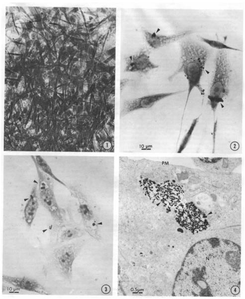
Figure 2-1 demonstrates the Ni3S2 induced morphological transformation in SHE cells. Cells exhibit disordered growth and criss-crossing. These disordered cells grow in soft agar and form tumors in athymic nude mice. Figure 2-2 is a light microscope photograph of SHE cells with arrows indicating the location of endocytized Ni3S2 particles which appear as black dots. Figure 2-3 is a light microscope photograph of CHO cells that have endocytized Ni3S2 particles with arrows indicating the location of the particles which appear as black dots are visibly encapsulated in a vesicle. Figure 2-4 is an electron micrograph of a CHO cell with endocytized Ni3S2 particles contained in a vesicle. Images from Costa and Mollenhauer 1980b. Reprinted with the permission of Cancer Research.

