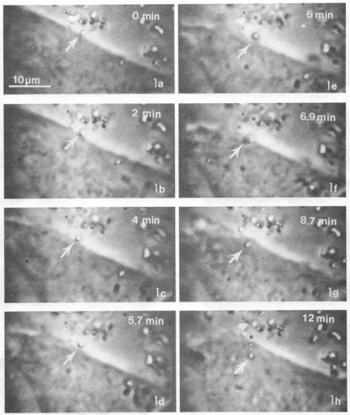
Figure 3.
Image of Cells Endocytizing Particles
Endocytosis of crystalline Ni3S2 particles. Images were recorded on videotape at 18/1 time lapse and photographs of the film were taken with Polaroid film. The total time lapse from images 3A-3H is 12 minutes. Ni3S2 particles appear white under phase contrast in these images. In 3a (0min) two particles are bound to the CHO cell surface. In panels A (0 min), E (6 min), and H (12 min) the particle indicated by the white long arrow is endocytized over the time course while the particle indicated by the white short arrow remains affixed to the cell surface.
Images from Evans et al. 1982. Reprinted with permission of Cancer Research.

