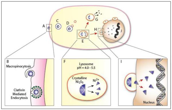
Figure 4.
Proposed Model of Particulate Nickel Uptake and Intracellular Distribution
The nickel particle, crystalline Ni3S2 affixes at the cell surface (A). We hypothesize that the particle enters via macropinocytosis and/or clathrin mediated endocytosis - where the different forms of uptake may be related to the size of the particle (B). In macropinocytosis the membrane exhibits ruffling prior to uptake, a feature that was frequently observed during Ni3S2 endocytosis. In clathrin-mediated endocytosis the membrane undergoes a morphological change via invagination and forms a membrane pit. A number of proteins are involved in CME, pictured here are the clathrin proteins that stabilize the pit curvature and the dynamin that aggregates at the neck prior to scission. The endocytized particle moves via saltatory motion towards the nucleus inside some form of vesicle (the specific form will vary in accordance with the form of endocytosis - macropinosome or clathrin coated vesicle) (C). Lysosomes then interact with the vesicle in a process of lysosomal attack (D). These interactions often lead to lysosomal fusion (E). Once fused, the pH of the vesicle may be altered through proton pumps leading to acidification of the vesicle and the dissolution of the crystalline Ni3S2 particle. This process produces high concentrations of Ni2+ (F). In some cases the Ni2+ may exit the vesicle into the cytoplasm where they can interact with biomolecules (G); while in other cases the vesicles will continue to travel towards the nucleus where they will aggregate at the nuclear membrane (H). Those aggregated at the membrane will promote the transfer of Ni2+ ions into the nucleus (I). The presence of Ni2+ can lead to multiple effects including DNA condensation and the modification of epigenetic marks including increased DNA methylation and loss of histone acetylation in H2A, H2B, H3, and H4 as well as an increase in H3K9 dimethylation, H3K4 trimethylation and ubiquitylation of H2A and H2B.

