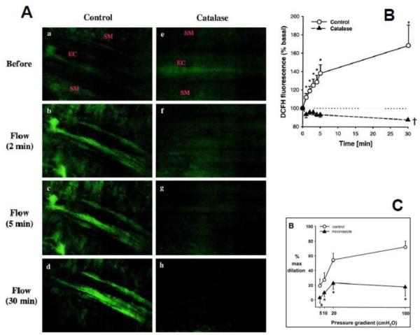
Figure 1. Hydrogen peroxide and epoxyeicosatrienoic acids are necessary for flow-mediated dilation of human coronary arterioles from patients with coronary disease.
Representative images of DCFH fluorescence (A) in an human coronary arteriole before (a and e) and after exposure to shear stress (b through d and f through h) in the absence or presence of catalase (1000 U/mL). a through d, Shear stress induces an increase in fluorescence intensity in the endothelial cell layer (EC) but not in the VSMC layer (SM). e through h, Increase in fluorescence to shear stress was not observed in vessels treated with catalase. Summary data (B) shows that shear stress induced endothelial H2O2 production occurred in a time-dependent manner (*P<0.05 vs baseline, n=5). Treatment with catalase completely abolished H2O2 production (P<0.05 vs control, n=4). Flow-induced dilation is also markedly inhibited by miconazole (C) or the selective epoxygenase inhibitor 17-ODYA. Reprinted from [45] and [46]. Reproduced with permission from the American Heart Association.

