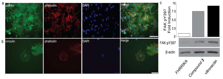
Figure 5.
Surfaces displaying the small molecule 3, which binds the αvβ3 integrin, activate signaling. a–b) M21 cells cultured on a surface presenting 3 were stained for paxillin (a, green) or for vinculin (b, green) and counterstained with phalloidin (red) and DAPI (blue). Scale bars, 100 μm (a) 50 μm (b). c) Histogram depicting change in focal adhesion kinase (FAK) phosphorylation (pY397) for cells cultured on the indicated surfaces; values were normalized to β-actin levels. Immunoblot analysis for cells cultured on indicated surfaces using a phospho-specific antibody against FAK pY397 and an antibody against β-actin.

