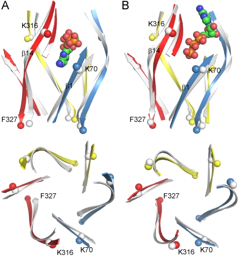
Fig. 5.
ATP-induced conformational changes of zfP2X4R in MD simulations. (A) ATP in proximal orientation. (B) ATP in distal orientation. β1 and β14 are shown in both side view (Upper) and top view (Lower). One ATP molecule and β13 in the top front left subunit are also shown in the side view. The β-strands in the resting state are shown in gray as reference. K70 and K316 are shown as spheres to highlight the closure between β1 and β14 at the top; F327 is shown as spheres to indicate whether the β14 C termini move apart.

