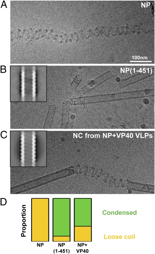
Fig. 3.
Minimum assembly component of the EBOV NC. (A) CryoEM image of purified full-length EBOV NP. Protein density is black. (B) Image of purified NP(1-451). Inset: 2D average of extracted helical segments. Width of box 720 nm, protein density white. (C) Corresponding images of the NC helix purified from NP+VP40 VLPs. (D) Comparison of proportion of condensed helices (green) and loose coils (yellow) observed in the three samples. Data values are in Table S1.

