Fig. 4.
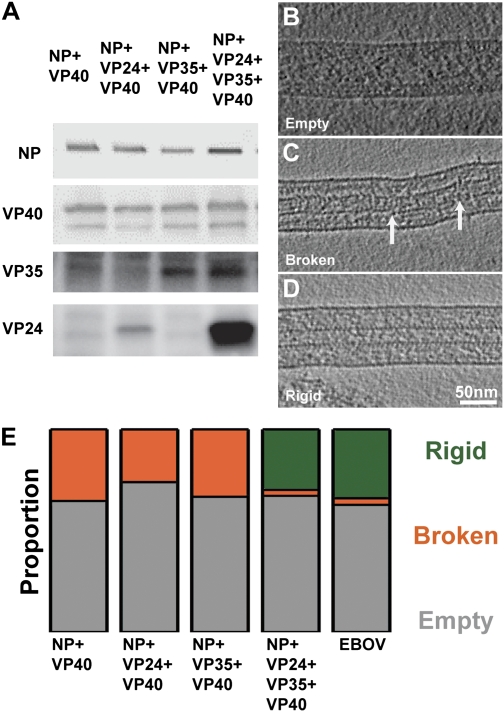
Protein recruitment and formation of a rigid NC. (A) Detection of viral proteins in respective VLPs. Purified VLPs were collected, and Western blot analysis using rabbit anti-NP, -40, -35, and -24 antibodies was performed. (B) A tomographic slice through an empty VLP. Protein density is black. (C) Slice through a VLP with a broken NC. Points of breakages in the NC helix have been highlighted with white arrows. (D) A VLP with a rigid NC. (E) Proportion of particles observed with a rigid NC (dark green), with an overall broken NC (orange), and without an NC (gray) in different samples. Data values are in Table S2.

