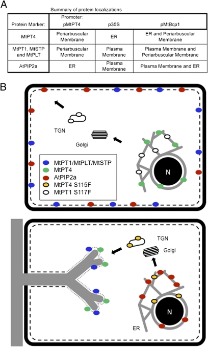
Fig. 6.
Summary and model for phosphate transporter secretion during arbuscule development. (A) Table summarizing membrane localizations and illustrating that proteins expressed from the MtBcp1 promoter show localization patterns that combine those obtained from MtPT4 and p35S promoters. (B) Proposed model derived from data obtained through analysis of protein-GFP fusions expressed either from their native promoters and/or expressed ectopically from promoters that show differential activity during arbuscule formation. The cartoon depicts an uninfected cell (Upper) and a cortical cell harboring a developing arbuscule (Lower). In an uninfected cell, MtPT1, MtSTP, MtPLT, and AtPIP2a are secreted to the plasma membrane (black dashed line), whereas MtPT4 is retained in the ER. The MtPT1S117F mutant protein is retained in the ER and likely in the TGN. During arbuscule development (Lower), newly synthesized MtPT4, MtPT1, MtSTP, and MtPLT, expressed from an arbuscule-specific promoter, are secreted to the periarbuscular membrane (gray dotted line), whereas AtPIP2a is retained in the ER. The mutant protein MtPT4S115F is retained in the ER and likely in the TGN.

