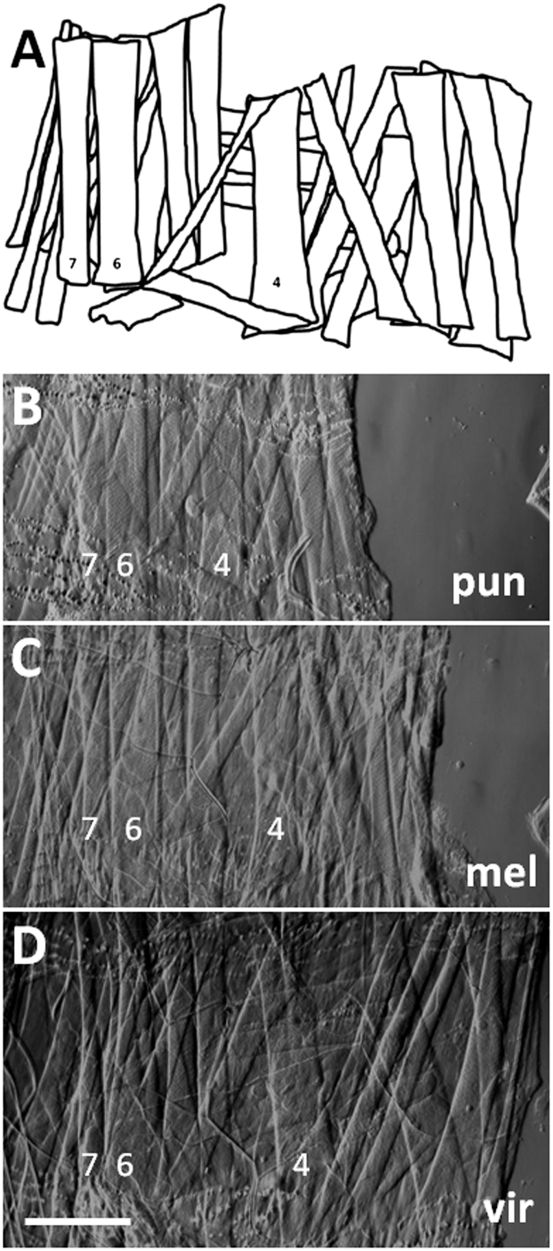
Fig. 1.
(A–D) Larval body wall musculature is precisely conserved among Drosophila species. Bright-field images of dissected third-instar larvae from D. punjabiensis (pun), D. melanogaster (mel), and D. virilis (vir) show musculature in right hemisegment 2. (A) Diagram illustrates the complete pattern of musculature in each of these species. All of these muscles cannot be seen in the microscopic images, which were focused on the most superficial muscle layers. For orientation, muscles 4, 6, and 7 are labeled. This analysis focused on the NMJs that form on muscle 4. (Scale bar, 200 μm.)

