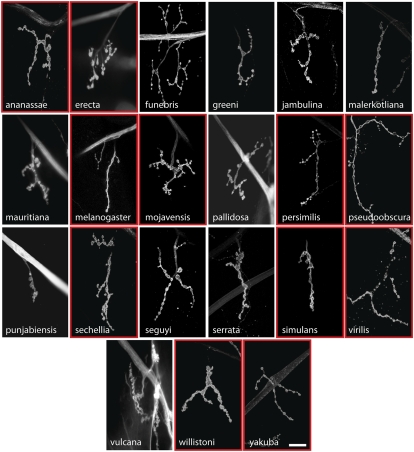
Fig. 2.
Larval NMJs show extensive morphological variation in different species of Drosophila. Third-instar larvae of each species were dissected and stained with FITC-conjugated anti-HRP. Representative confocal images of NMJ4 in abdominal segment 2 are shown. There is substantial variation in NMJ morphology among these species with respect to overall geometry, number of boutons, and number of branch points. The 11 species enclosed by red boxes are those whose genome has been completely sequenced and whose precise phylogenetic relationships have been ascertained. (Scale bar, 20 μm.)

