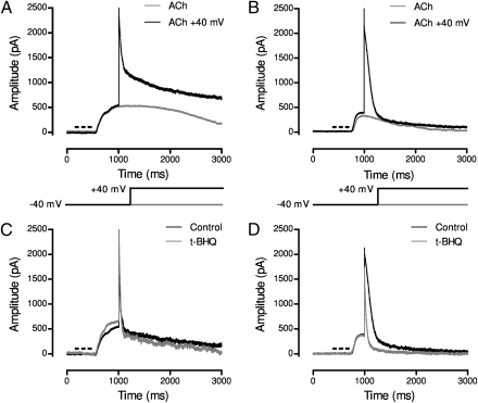
Fig. 4.
ACh-evoked potassium current in chicken and rat hair cells. (A) A puff of ACh (100 μM for 300 ms) caused an outward current at −40 mV lasting approximately 2 s in rat inner hair cells (gray). When the membrane potential was stepped to +40 mV, the initially large outward current rapidly decayed (black) to a level dominated by a voltage-dependent outward current. (B) ACh-evoked membrane current in a chicken hair cell lasting approximately 2 s at −40 mV (gray). With a step to +40 mV, the initially large outward current decayed, albeit relatively slowly, to a level reflecting the small voltage-dependent outward current (black). (C) ACh-evoked “difference current” after subtracting membrane current in the absence of ACh in a rat inner hair cell under control conditions (black) and after exposure to the SERCA inhibitor t-BHQ (gray). (D) Same as in C, but for a chicken short hair cell.

