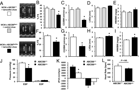
Fig. 2.
Cardiac function is compromised in ABCB8 KO mice. (A) M-mode echocardiography images of control and ABCB8 KO mice. Two sets of control animals are used in these studies: MCM × ABCB8+/+ mice treated with tamoxifen chow (white bars) and MCM × ABCB8f/+ mice treated with regular chow (gray bars). (B–E) Control and ABCB8 KO mice were analyzed by echocardiography 4 wk after tamoxifen treatment. Both percentage of FS (measure of ventricular contractility) and CO/BW (the amount of blood pumped by the heart) are significantly reduced, whereas LVDd (measure of chamber dilation) and HW/BW (measure of cardiac size) are not different in ABCB8 KO mice compared with control (n = 6). (F–I) Echocardiographic analysis of control and ABCB8 KO mice at a delayed phase (8 wk after tamoxifen treatment; n = 6). (J) Invasive hemodynamic measurements 8 wk after tamoxifen treatment. (K) dP/dtmax (contractility) and dp/dtmin (relaxation) in WT and ABCB8Δ/Δ mice 8 wk after tamoxifen treatment. (L) Heart rate during hemodynamic measurements in WT and KO mice (n = 6). Data are presented as mean ± SEM (*P < 0.05).

