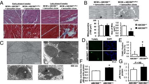
Fig. 3.
ABCB8 KO hearts display structural abnormalities. (A) Histological analysis of hearts from control (MCM × ABCB8+/+ treated with tamoxifen) and ABCB8Δ/Δ mice 4 and 8 wk after tamoxifen treatment. Upper: H&E eosin staining. Lower: Masson trichrome staining. Blue staining represents cardiac fibrosis. (B) Representative EM images of hearts from ABCB8+/+ and ABCB8Δ/Δ mice. Arrows indicate mitochondria. The graph below shows quantification of mitochondrial number and size (n = 5). (C) Representative EM images of hearts from ABCB8+/+ and ABCB8Δ/Δ mice. Hearts from ABCB8+/+ mice show well aligned mitochondria with clearly distinguishable cristae. Mitochondria from ABCB8Δ/Δ mouse hearts show accumulation of electron-dense material (asterisks), disruption of cristae, and deformed morphology. (D) LV myocardial sections from ABCB8+/+ and ABCB8Δ/Δ mice subjected to TUNEL and DAPI (nuclei) staining after 4 wk reveal higher cell death in ABCB8Δ/Δ mice. (E) Quantitative analysis of five fields as shown in D (n = 6). (F) Mitochondrial nonheme iron levels are higher in ABCB8 KO in the heart (n = 5). (G) The levels of mitochondrial heme are not altered in the hearts of ABCB8Δ/Δ mice (n = 3). Data are presented as mean ± SEM (*P < 0.05).

