
Fig. 4.
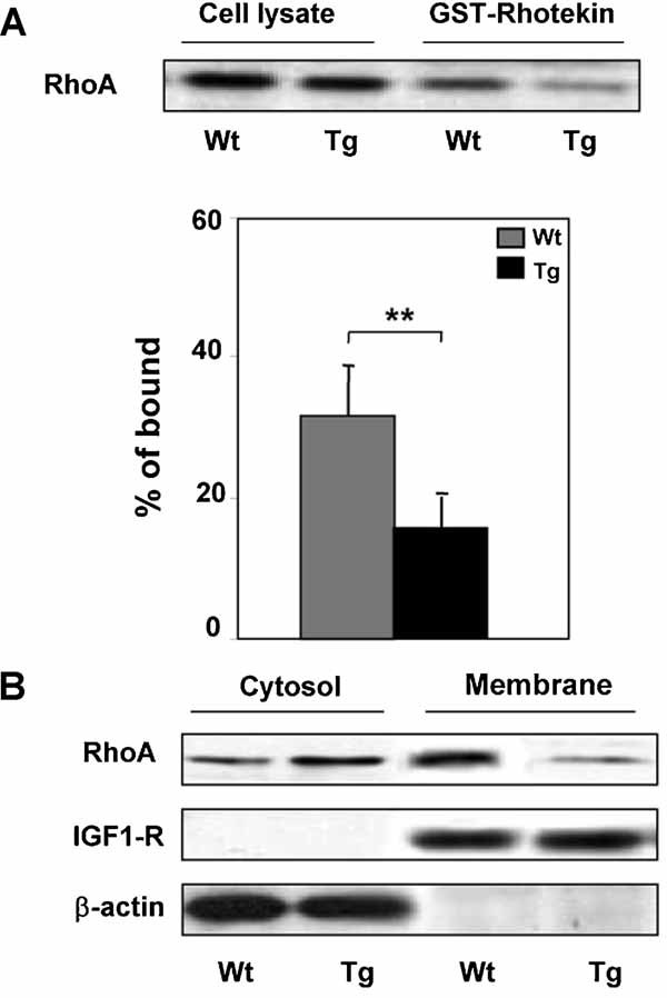
RhoA activity and localization in fibroblasts from Wt and TgPed fibroblasts. A: Cell lysates were obtained from Wt and TgPED fibroblasts as described in “Materials and methods” Section. Pull-down assays with GST-Rhotekin were performed and analyzed by Western blot. Blots were revealed by ECL and autoradiography. Densitometry for pulled-down RhoA-GTP was normalized to the amount of total RhoA. The results are presented as percentage of bound respect to total level of protein. Data are mean of four independent experiments. Asterisks denote statistically significant differences (**P < 0.01). B: Cytosolic and membrane fractions of Wt and TgPED fibroblasts were obtained as described in “Materials and methods” Section, subjected to SDS–PAGE and immunoblotted with anti-RhoA, β-actin and IGF-1 receptor β-subunit antibodies, as indicated. Blots were revealed by ECL and autoradiography. The blots shown are representative of four independent experiments.