
Fig. 6.
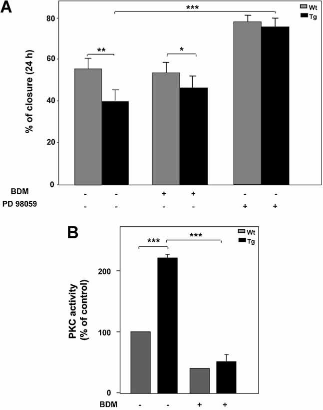
Effect of PKC and ERK inhibitors on in vitro wound healing. A: Confluent monolayers of fibroblasts from Wt and TgPED mice were subjected to scratch assays, as described in “Materials and methods” Section, and in the legend of Figure 1. The scratch assays were performed in culture medium alone or added with 5 µM BDM or 30 µM PD98059, as indicated. Healing was calculated as described in “Materials and methods” Section. Bars represent the mean ± SD of triplicate determination in four independent experiments. Asterisks denote statistically significant differences (*P < 0.05; **P < 0.01; ***P < 0.001). B: PKC activity was assayed as described in “Materials and methods” Section. PKC activity is expressed as percentage over control activity. Bars represent the mean ± SD of data from four independent experiments. Asterisks denote statistically significant differences (***P < 0.001)