
Fig. 9.
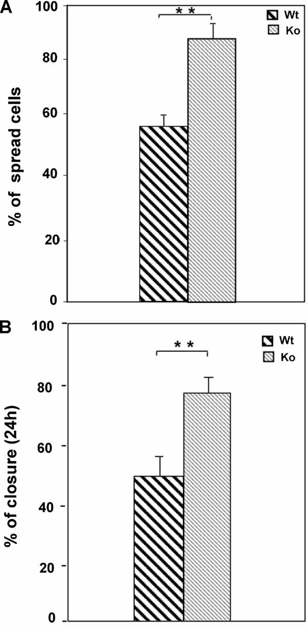
Cytoplasmic spreading and in vitro wound healing in ped/pea-15 null fibroblasts. A: Fibroblasts from Wt and ped/pea-15 null mice were subjected to spreading assays. After 3 h, cytoplasmic spreading on fibronectin was quantified by light microscope after staining with crystal violet as described in “Materials and methods” Section. Bars represent the mean ± SD of three independent determinations in triplicate. Asterisks denote statistically significant differences (**P < 0.01). B: Confluent monolayers of fibroblasts from Wt and ped/pea-15 null mice were subjected to scratch assays, as described in “Materials and methods” Section. Healing was calculated as described in “Materials and methods” Section. Bars represent the mean ± SD of triplicate determination in four independent experiments. Asterisks denote statistically significant differences (**P < 0.01).