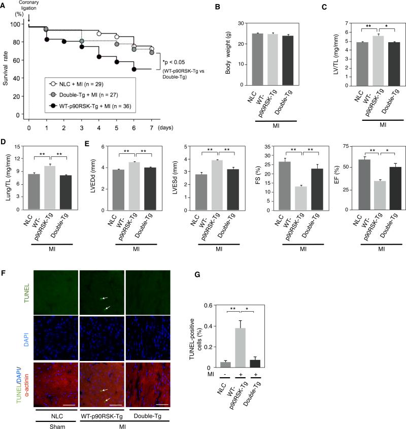
Figure 7. ERK5 activation prevented the exacerbation of LV dysfunction after MI in WT-p90RSK-Tg mice.
(A) Kaplan-Meier survival analysis in NLC, WT-p90RSK-Tg, and Double-Tg-WT-p90RSK-Tg/CA-MEK5α-Tg (Double-Tg) after MI. Survival rate in NLC (n=29), WT-p90RSK-Tg (n=36), and Double-Tg mice (n=27) after MI are plotted. Overall survival was significantly higher in Double-Tg compared to WT-p90RSK-Tg mice. * p<0.05 compared to WT-p90RSK-Tg group. (B) Body weight one week after MI in NLC, WT-p90RSK-Tg, and Double-Tg mice. (mean±S.D., n=17-19) (C, D) LV weight/TL (C) and lung weight/TL (D) after MI in NLC, WT-p90RSK-Tg, and Double-Tg mice. (* p<0.05, ** p<0.01, mean±S.D., n=17-19) (E) Echocardiographic data obtained after MI in NLC, WT-p90RSK-Tg, and Double-Tg mice. LVEDd, left ventricular end-diastolic dimension; LVESd, left ventricular endsystolic dimension; TL, Tibial length; FS (fractional shortening); EF (ejection fraction). (* p<0.05, ** p<0.01, mean±S.D., n=17-19) (F) Cardiomyocyte apoptosis in the remote area was increased in WT-p90RSK-Tg mouse hearts, which was inhibited in Double-Tg mice. TUNEL-positive cardiomyocytes were counted in the remote area of each mouse heart as described in Fig.6D. Representative pictures of TUNEL (top), DAPI (middle), and α-actinin merged with TUNEL and DAPI staining (bottom) of the remote area from NLC, WT-p90RSK-Tg, and Double-Tg mice subjected to MI or sham operation. 40X objective lens. Scale bars, 40 μm (G) A bar graph showing TUNEL-positive cells (%) in various animals. (** p<0.01, *p<0.05, mean± S.D., n=3).

