
Figure 4. Proliferating cell survival and neuronal differentiation.
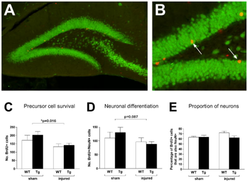
BrdU was administered beginning two weeks postinjury for seven consecutive days. Animals were euthanized three weeks after the final injection (six weeks postinjury) and assessed for proliferating cell survival (BrdU+, red) and neuronal differentiation (double-labelling with NeuN, green) within the dentate gyrus. The majority of BrdU+ cells were located in the subgranular zone (A, GPx Tg sham) where double-labeled cells could also be found (B, arrows). As with Ki-67, the interaction between genotype and injury had no effect on proliferating cell survival (p=0.56) but injury resulted in a significant reduction (p=0.016, C). A similar pattern was seen with neuronal differentiation, with injury trending towards an effect (p=0.087, D). Thus, while injury reduces proliferating cell survival and possibly neuronal differentiation, GPx overexpression is unable to rescue those changes. No differences were seen in the percentage of BrdU+ cells that differentiated into neurons (E). Scale bars: A 100μm; B 50μm.