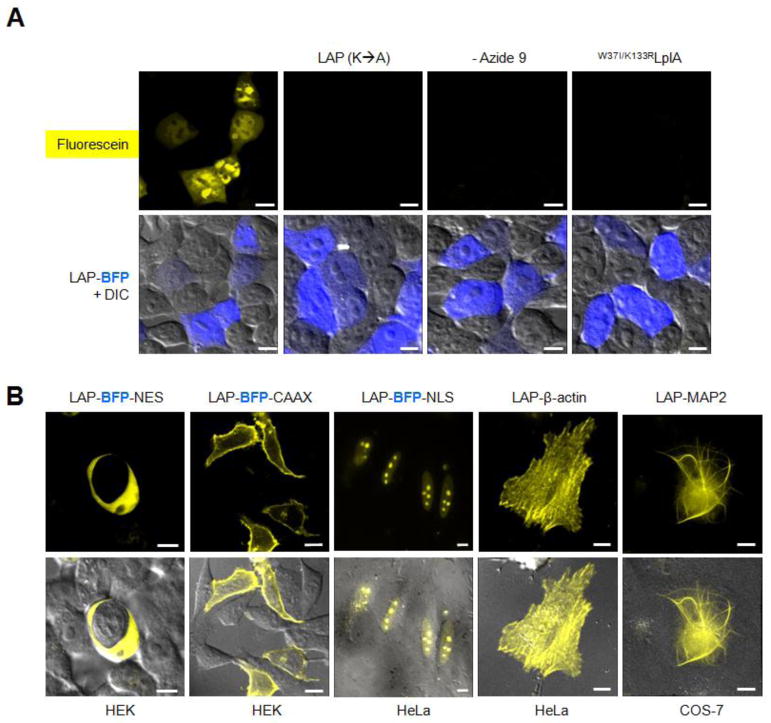
Figure 4.
Intracellular protein labeling with azide 9 ligase and ADIBO-fluorescein. (A) HEK cells co-expressing W37ILplA and LAP-BFP were labeled with azide 9 and ADIBO-fluorescein as in Figure 3A, then imaged live. Negative controls are shown with an alanine mutation in LAP, azide 9 omitted, and a catalytically inactive mutant of LplA (last column). (B) ADIBO-fluorescein labeling of three localized LAP-BFP fusions, LAP-β-actin, and LAP-MAP2 (microtubule-associated protein 2). Labeling in the cell type indicated beneath each image was performed as in Figure 3A, except that for LAP-β-actin and LAP-MAP2, azide 9 was incubated for 2 hr, and washed for 1.5 hr before fluorophore addition. NES = nuclear export sequence; CAAX = prenylation tag; NLS = nuclear localization sequence. All scale bars, 10 μm.

