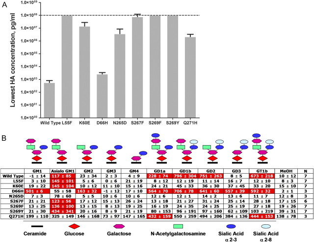
Figure 2.
JCV VLPs carrying PML-associated mutations lose the ability to hemagglutinate red blood cells (RBCs) and bind sialylated gangliosides. (A) Ability of mutant VLPs to hemagglutinate red blood cells from different blood groups. RBCs were incubated with serial 2-fold dilutions of various VLPs, starting from 100 μg/mL. Minimum hemagglutination (HA) concentration is the lowest concentration of VLP that still agglutinated RBCs. HA results were examined by visual inspection. All HA reactions were conducted in duplicates. Mean ± SD for the minimum HA concentration is calculated based on the results obtained from hemagglutination of 4 to 7 different blood group donor RBCs (A, B, O, and AB). VLPs displaying the minimum HA concentration of 100 μg/mL (dotted line) did not cause any hemagglutination at this concentration (ie, highest VLP concentration tested). (B) Specificity of JC VLPs for sialylated gangliosides. VLPs binding to an array of gangliosides were detected with a 2-step process involving the detection of VLPs bound to a ganglioside with VP1-specific murine antibodies and anti-murine IgG HRP–labeled antibodies followed by development with TMB substrate. Numbers represent percent increase in the optical density obtained with the specific VLP relative to that obtained without VLP (ie, background) and were calculated as follows: % specific VLP binding = 100%*(OD450(with VLP)–OD450(without VLP))/OD450(without VLP). Statistically significant (P < .05) interaction between a VLP and ganglioside in comparison to the background interaction between the VLP and MeOH-treated well is denoted by a red cell background. Results are depicted as a mean ± SD binding for each VLP to each of the gangliosides or to the control for several independent experiments conducted; N denotes number of independent measurements for each VLP and all gangliosides. Schematic structure of ganglioside is shown to reveal core binding structure bound by various VLPs.
NOTE. JCV indicates JC virus; VLPs, virus-like particles; PML, progressive multifocal leukoencephalopathy; VP1, viral protein 1; TMB, tetramethylbenzidine.

