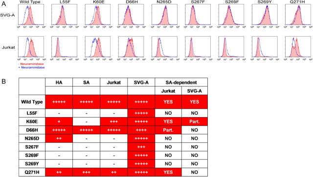
Figure 4.
Binding of JCV VLPs carrying PML-associated mutations to glial cells is neuraminidase treatment–insensitive. (A) SVG-A glial cells (top row) or Jurkat lymphoid cells (bottom row) were first either pretreated with neuraminidase (blue line) for 60 min at 37°C to remove all terminal α2–3–, α2–6–, and α2-8–linked sialic acid residues or mock-treated (red line) followed by incubation at 4°C with the indicated VLP and staining with the detection antibodies as described in the previous figure. One representative experiment out of 4 conducted is shown. Staining of the cells with Sambucus nigra agglutinin (SNA) lectin and Maackia amurensis lectin II (MAL II) proved neuraminidase treatment effectiveness in sialic acid removal (data not shown). (B) Sialic acid dependence as compared with VLP binding by various assays. The degree of binding was evaluated as a percent of wild-type VLP binding value in the assay (Figures 2 and 3) and were calculated as follows: 100%*(VLP VALUE – BASELINE VALUE)/(WT VALUE – BASELINE VALUE); hemagglutination assay values were LOG10 transformed first. Values were ranked accordingly: “–” < 10; 10 < “+” < 20; 20 < “++” < 40; 40 < “+++” < 60; 60 < “++++” < 80; and “+++++” > 80.
NOTE. JCV indicates JC virus; VLPs, virus-like particles; PML, progressive multifocal leukoencephalopathy; HA, hemagglutination; SA, sialic acid binding; HRPTEC, human renal proximal tubular epithelial cells; astrocytes, primary human astrocytes. Dependence on sialic acid binding is denoted as NO, no dependence; or Part., partial dependence. Positive values are denoted by red cell backgrounds.

